


PNNL Imaging Method Maps How Leafcutter Ant Fungus Gardens Break Down Plant

Researchers at Pacific Northwest National Laboratory (PNNL) developed an imaging method, metabolome informed proteome imaging (MIPI), that visualises plant breakdown inside a leafcutter ant fungus garden, and published it in Nature Chemical Biology.
The method scans thin garden sections with a laser to locate metabolites, plant polymers and enzymes, and the team used it to identify the fungus Leucoagaricus gongylophorus as the primary degrader of plant material.
PNNL says the findings could inform future biofuels and bioproducts, but the source material shows no conversion process or commercial application yet.
A Garden That Digests Leaves
Researchers at Pacific Northwest National Laboratory have built an imaging method to show where and how the fungus gardens of leafcutter ants break down plant material, with bioproduct development in mind. The technique, metabolome informed proteome imaging (MIPI), was developed by Kristin Burnum-Johnson, science group leader for Functional and Systems Biology, and colleagues. The work was part of her US Department of Energy (DOE) Early Career award.
Scientists have spent decades learning to degrade plant matter into bio-based fuels, detergents, nutritional supplements and plastics. Lignin, a primary component of plant cell walls, remains hard to break down affordably without adding pollutants back into the environment, and it can end up as waste. Companies already turning plant residues into ingredients, as in fungal fermentation of carrot waste, work on the same underlying challenge.
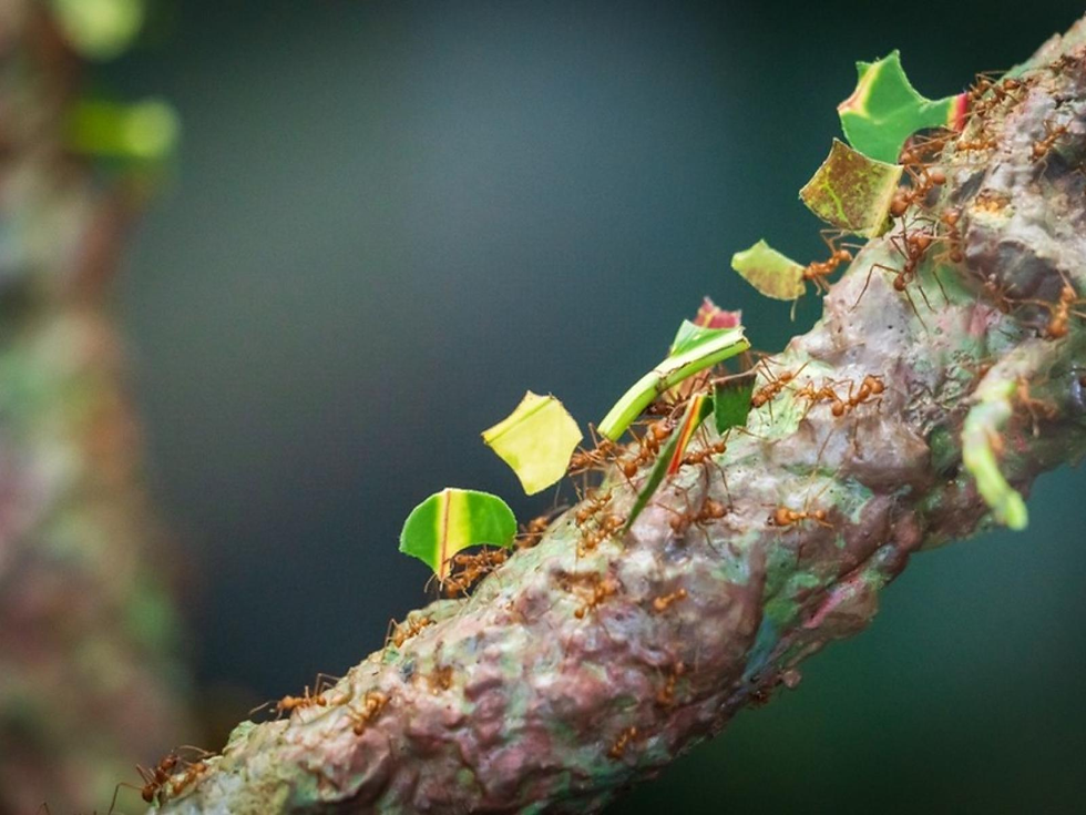
Leafcutter ants offer a natural example. They cultivate Leucoagaricus gongylophorus on fresh leaves in specialised underground structures. These become fungal gardens whose leftover components are consumed by other organisms in the system. Resident bacteria support the garden by producing amino acids and vitamins. According to PNNL, working out every component and reaction involved had remained a significant challenge, because the plants, fungus, ants and bacteria do not each stay in one place or perform one task.
Imaging the Breakdown at Microscale
The team used a high-powered laser to scan sections of fungal garden 12 microns thick, roughly the width of plastic cling film. The scans located metabolites, the remnant products of plant degradation. They also located plant polymers, including cellulose, xylan and lignin. Where these overlapped, they marked hot spots of plant breakdown.
The researchers then examined those regions for enzymes, which start biochemical reactions, and used enzyme type and location to work out which microbes were involved. PNNL reports that this confirmed the fungus as the primary degrader of the plant material. It also found that the bacteria transform previously digested plant polymers into metabolites used as vitamins and amino acids. These, according to the team, accelerate fungal growth and plant degradation.

Burnum-Johnson argued that conventional bulk measurements average out the signal, diluting the chemical reactions of interest and often making them undetectable. Marija Velickovic, the paper's lead author, performed the laboratory work, which included scanning samples and identifying hot spots of lignocellulose degradation. The research was funded by the DOE Office of Science and used resources at the Environmental Molecular Sciences Laboratory, a DOE user facility at PNNL.
What Earlier Proteomics Showed
The fungus's flexibility was examined in an earlier study that also used proteomics, the large-scale analysis of proteins. Researchers excavated five Atta cephalotes colonies near the Smithsonian Tropical Research Institute's Gamboa station in Panama and split the fungus chambers into sub-colonies. These were fed leaves, Hibiscus rosa-sinensis flowers, oats or a mixture of all three for 15 days.
The analysis identified 1,766 fungal proteins, including 161 putative biomass-degrading enzymes. Gardens fed leaves or flowers produced more proteins predicted to break down cellulose, namely endoglucanase, exoglucanase and β-glucosidase. The mixed diet resembled the oat-only diet, which the authors interpret as the fungus preferentially digesting simpler substrates when they are available.

That study's treatment of bacteria was more cautious. Only 44 unique bacterial peptides were detected, about 1% of unique peptides, and three bacterial proteins differed between treatments. The authors suggest this may reflect a large difference in fungal and bacterial biomass, or possibly a more limited bacterial role. PNNL's later work, by contrast, assigns the bacteria a role in supplying vitamins and amino acids. The sources do not reconcile the two framings; the studies used different methods and questions.
Limitations and the Road Ahead
PNNL's account gives no quantitative performance data, and the claim that the findings can be applied to biofuels and bioproducts is a stated expectation, not a demonstrated process. For developers working on lignocellulose conversion, the sources offer a map of how a natural system divides the labour. They do not show that this translates into cheaper or cleaner processing.
The earlier Panama study has its own constraints. It used laboratory sub-colonies, and 16 samples were analysed (four per treatment). The bacterial findings rested on single peptides. Some sub-colonies failed during the experiment, and oat-only sub-colonies had significantly lower survivorship. The authors said colony health was not the study's focus and offered only a hypothesis about missing micronutrients.
PNNL says the team's next step is to study how fungal communities respond and protect themselves amid disturbances. Whether that, or the MIPI imaging itself, leads to industrial enzyme discovery or bioprocess design remains an open question.


